
A new device in CivMin’s repertoire of high-tech machines allows researchers to peer inside solid objects. Using X-ray and computed tomography (CT) scans, the new system enables detailed 3D imaging in a non-destructive manner, literally allowing the unseen to become seen, and displayed upon a screen, with incredible detail.
This technology is of benefit not simply at CivMin, predominantly for the study of layering and fractures within rock specimens, but also for other departments and faculties at U of T and beyond. Imagine peering into solid objects without damaging the exterior or opening a sealed container, such as a very old manuscript with pages fused together, embedded objects within a block of ice, or even remains contained in ancient, unopened pottery.
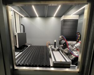
X-ray micro computed tomography (micro-CT) is a non-destructive imaging technique capable of characterizing the internal microstructure of materials in three dimensions at micron-level resolution. The concept of this technique is based on the sectioning of an object with many radiographs captured at different angles. These radiographs are formed by the partial absorption of the X-ray energy into the object and with the rest passing through, corresponding to density and material thickness, consequently revealing the internal structure.
In the context of research within Grasselli’s Geomechanics Group (GGG), the micro-CT is a state-of-the-art tool which will elevate the quality of the research by enabling the 3D investigation of materials before, after, and even during experiments. Using the data, a 3D digital image of the rock can be produced allowing for the physical study of macro-features like planes of heterogeneity and disconformities to more microscopic applications such as micro-cracks and pore network. Advanced machine learning techniques and artificial intelligence methods can be leveraged on the image volume to provide fracture morphology characteristics, as well as those of the pore spaces such as its size distribution, interconnections, and tortuosity, as well as the segmentation of the various phases present in the sample or identify connectivity pathways.
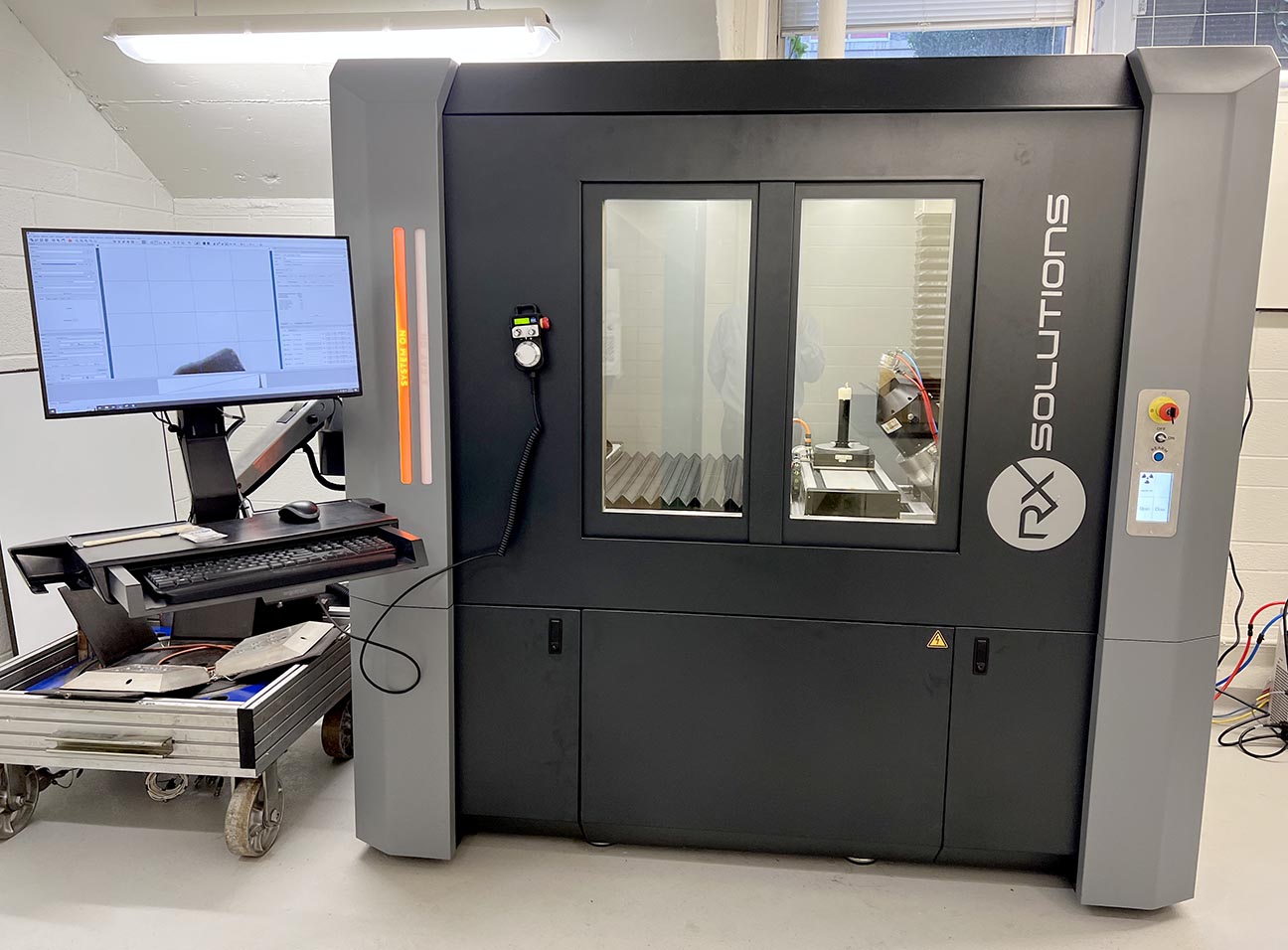
Funded through the Canada Foundation for Innovation Evans Leaders Fund (CFI-JELF), Prof. Giovanni Grasselli and the Transparent Lab have purchased the RX Solutions EasyTom 230 Micro-CT X-Ray System. The RX Solutions EasyTom Micro-CT is an industrial high-energy CT System capable of imaging the internal structure of objects in a fast, non-destructive manner. It allows researchers to accurately examine internal and external structures to address the most challenging 3D applications ranging from material research to industrial applications in research and development, quality assurance, and production.
Watch a 3D view of a scan using a Micro-CT X-Ray System
Credit: Ms. Mei Li (PhD Candidate) and Mr. Earl Magsipoc (PhD Candidate)
The EasyTom Micro-CT can generate image volumes with voxel sizes down to 4 µm with a detection area of 320 mm in diameter and 530 mm in height and can image payloads with a mass of up to 20 kg. It is equipped with six motorized axes to position the sample and detector with up to 590 mm displacement stroke. The EasyTom has a tube voltage range of 0-220 kV and a tube current range of 0-1000 µA, with a maximum power of 200 W. From the set of vertically acquired radiographs, a mathematical algorithm integrated in the EasyTom X-Act software suite helps to streamline acquisition, enhances reconstruction with advanced corrective algorithms, and provides an automated workflow all the way through scanning, reconstruction, and inspection steps.
(Photo by Phill Snel, CivMin)
The current research work undertaken within the research group and benefits gained from the acquired micro-CT align with the current trajectory of the geoenergy sector, such as the National Resources Council (NRC) Canada Advanced Clean Energy program: Hydrogen. Hydrogen storage in subsurface salt-caverns is going to be a key aspect for the energy transition in Canada. Through direct X-ray micro-CT imaging, it will be possible to improve our understanding of the complex multiphysics phenomena associated to H2 rapid cycle injections in salt caverns necessary to develop sound legislation and safety protocols.
Within the scientific community, and that of the University of Toronto, the benefits are endless and extend way beyond rock mechanics. The EasyTom Micro-CT at the Transparent Lab will support cutting edge research within a variety of fields, for example:
- Dental – Medical Science: Track the development and remodelling of bone structure to understand bone/dental implant materials (e.g., titanium, dental epoxy) as well as impacts of gut microbiome on bone structure throughout life.
- Fluid Mechanics – Water Treatment: Image phase transition processes in ice as well as understanding multiphase and biogeochemical processes for clean energy applications, carbon sequestration, and various groundwater remediation technologies.
- Mining – Environment: Evaluating changes in soil pore structure in wetlands and bioretention cells when subjected to freeze-thaw cycles, and quantifying the potential for accumulation of suspended solids and microplastics.
- History: Reveal the internal structures and identify writing and origins of invaluable and fragile manuscripts to study the global evolution of book technology and characterize construction materials (e.g., types of paper, wood, ink etc.).
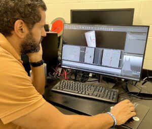
By Aly Abdelaziz (PhD Candidate) with Phill Snel